×

![]() Overview | Causes | Symptoms | Diagnosis | Treatment | FAQ
Overview | Causes | Symptoms | Diagnosis | Treatment | FAQ
Spondylolysis relates to instability of specific bones in the low back. It a very common cause of back pain, particularly in adolescents. Gymnasts who perform routines that bend and arch the back are often victims of spondylolysis or spondylolisthesis.
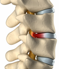
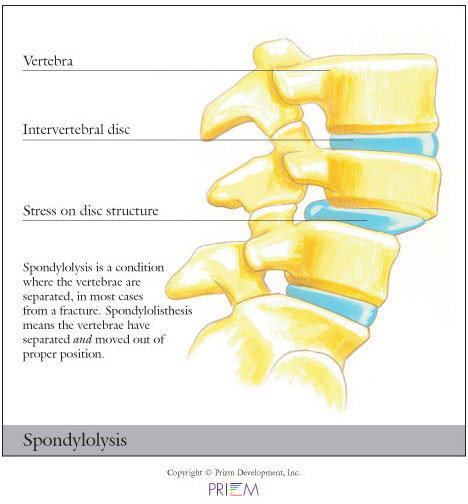
Spondylolisthesis and spondylolysis are caused by joint instability
in the low back. The rear part of spinal vertebrae has facet joints
that act as hinges, allowing our spines to twist and bend. Sometimes,
however, the posterior element can crack. Either from heredity or
wear and tear, part of the posterior element called the pars interarticularis
can crack, causing the vertebrae slip forward out of its correct
position. Spondylolysis occurs when the PARS hinge is cracked, but
the vertebrae is still in its correct position. Spondylolisthesis
occurs when the cracked PARS has allowed the vertebrae to slide forward
out of its correct position. If left untreated, spondylolysis can
lead to spondylolisthesis.
[Top]

Interestingly, in many cases, spondylolisthesis may have no symptoms,
so most people may not know they have it. Back pain is the most common
symptom, particularly in the lower back. This back pain may be mistaken
for a muscle strain. Muscle spasms that occur as a result of spondylolysis
may cause an overall feeling of stiffness in the back and may effect
posture.
[Top]
Outlined below are some of the diagnostic tools that your physician may use to gain insight into your condition and determine the best treatment plan for your condition.
Conservative treatments should always be considered first when treating
spondylolysis. Nonsurgical treatment methods include resting and
refraining from usual activities, taking anti-inflammatory medication,
and incorporating a stretching and strengthening program. While ligaments
and muscles can help hold the vertebrae in place, over time, surgery
may be necessary to install surgical instrumentation or bone grafts
that lock the vertebra in place so that it does not slide out of
position and damage the spinal nerves. Surgery may involve a fusion
and/or screws and rods.
[Top]
Those with a family history of spondylolysis or weak vertebrae are
more susceptible to developing the condition. Also, athletes involved
in activities that place a great deal of stress on the back, such as
football players and weight lifters, are at greater risk for fracturing
the vertebrae, encouraging slippage.
[Top]
We have made it easier to schedule appointments, click the button to fill out an online appointment request form or call our new central scheduling line 401-457-1500 to schedule an appointment.
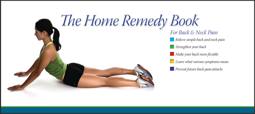
Have back or neck pain? Learn what causes symptoms and the home remedies that relieve pain. University Orthopedics mails out Home Remedy Books to residents throughout the New England area.
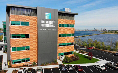
University Orthopedics in Providence, Rhode Island, is a regional referral center for patients with back and neck pain, joint pain, sports medicine problems, shoulder pain, hand problems, hip and knee pain, and foot and ankle injury. University Orthopedics utilizes fellowship-trained, sub-specialized physicians to treat a variety of orthopedic problems. Learn more.