×
Spinal column | Cervical spine | The spinal cord | Lumbar spine and sacrum | Healthy disc | Bone spur | Spondylolysis| Stenosis | Herniated & Bulging disc | Spine tumors | Degenerative disc | Muscles| Joints
To communicate with your back doctor, it helps to know the terms your physician might use to explain and describe your condition. Just as dentists use a number to identify each tooth, a spine doctor has a labeling system for each link on the chain that makes up the spinal column. To clear things up a bit, here is a "crash course" in spinal anatomy.
Click on image to enlarge
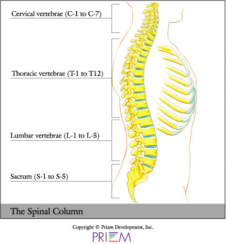
When viewed from the side, a healthy spinal column is slightly S-shaped. The top seven vertebrae are known as the cervical vertebrae, labeled C1 through C7.
The chest area contains the thoracic vertebrae, T-1 through T-12. The thoracic vertebrae do not rotate as much as the neck and low back. Consequently, this area of the spine is more stable and is generally less susceptible to injury. Relatively few back pain cases involve the T-level vertebrae.
Below the thoracic vertebrae are the five lumbar vertebrae, and below that is the sacrum. The lumbar vertebrae are labeled L1 to L5. This area is the most prone to injury, because it bears the most weight when you sit, stand, push, pull or lift.
Below the lumbar spine area is a series of fused bones known as the sacrum. At the bottom tip of the spinal column structure is the coccyx or the tailbone.
Each rounded vertebra body has pedicles and laminae, facet joints, and the bony transverse and spinous processes, which are the narrow, finger-like spikes pointing out from the sides and back of the vertebra.
Click on image to enlarge
This spinal column is held in place by surrounding muscles,
ligaments and tendons that act as supporting guy wires. When working
properly, the spine is able to bend and twist. When muscles and ligaments
weaken, problems arise in the stability of the spine. Muscles and ligaments
can strain, and discs and facet joints can be injured.
[top]
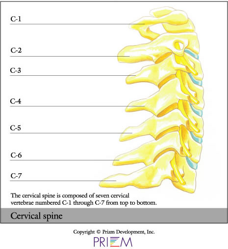
The cervical vertebrae make up the neck. Each vertebra in the cervical
region is labeled C-1 through C-7. The cervical vertebrae protect
the spinal cord, which attaches to the brain.
[top]
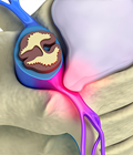
Within this column of vertebrae is the spinal cord, which travels from the brain stem down through the back. The spinal cord acts as our main electrical wiring system and is protected by the bony vertebrae. At every vertebrae level, there are nerve roots that branch off the spinal cord. When a disc herniates, it can crimp or pressure these nerve roots, which can cause excruciating pain that radiates into an arm or leg.
Nerve impingements in the cervical area can cause pain
to radiate into the shoulder and arm. When discs are injured in the
low back area, pain can radiate into the legs.
[top]
Click on image to enlarge
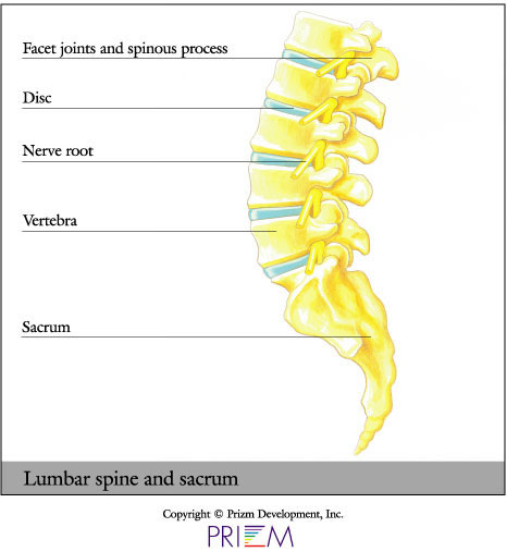
The lumbar area, or low back, contains L-1 through L-5, the largest, most sturdy group of vertebrae. Because it bears most of the body's weight when we sit, stand, push, pull, lift, and move, the lumbar section is considered the most injury-prone area of the spine. The spinal cord threads from the brain down through the spine and ends at about L-2, where it forms a bundle of nerves known as the cauda equina (Latin for 'horse's tail'). From the neck area to the coccyx are 31 pairs of nerve roots that exit the spinal canal and head for remote areas of the body through vertebral portals called foramina. At the base of L-5 is a solid mass of five fused bones called the sacrum (pronounced 'say-crum'). Finally, the spinal column ends at the coccyx (pronounced 'cock-six'), or tailbone, which is actually several small bones fused together.
Click on image to enlarge
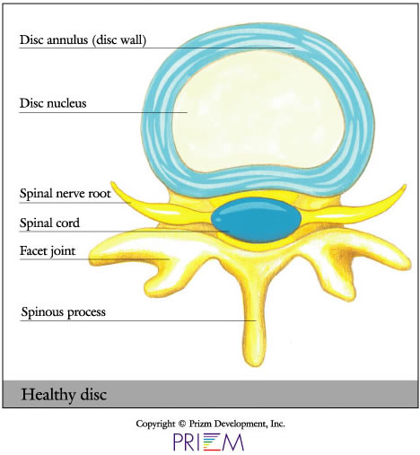
The spinal disc is like a jelly doughnut. The "jelly" of
the doughnut represents the "disc nucleus," and the material
that encases the "jelly" is called the "disc annulus." The
disc acts likes a rubber shock absorber between the vertebrae. The
facet joints act as hinges that allow for twisting and turning of the
spinal column. The spinal cord threads through from top to bottom like
a telephone wire system.
[top]
Click on image to enlarge
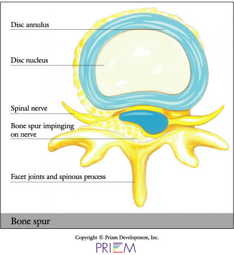
A
bone spur, or osteophyte, is a projection of bone that develops
and grows along the edge of joints. Bone spurs are fairly common
in people
over the age of 60. It is not the bone spur itself that is
the real problem; pain and inflammation begin to occur when the
bone spur
rubs against nerves and bones.
[top]
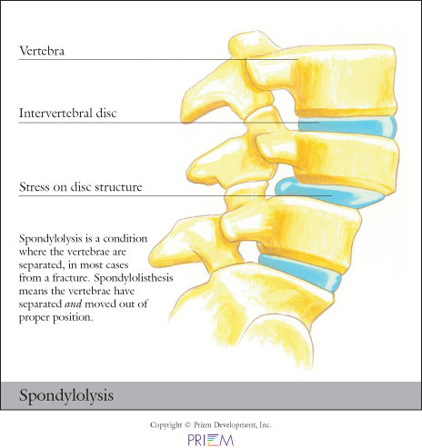
Spondylolysis relates to instability of specific bones in the low back.
It a very common cause of back pain, particularly in adolescents.
Gymnasts who perform routines that bend and arch the back are often
victims of spondylolysis or spondylolisthesis.
[top]
Click on image to enlarge
Stenosis is a condition that can develop as a person ages, particularly
in those over 50. It is characterized by a narrowing of the spinal
canal, which places pressure on the spinal cord and nerves, because
there is not enough room for them. It resembles placing a ring on
your finger. If the finger becomes injured or inflamed, the ring
constricts and causes pain. The pain caused by stenosis is typically
focused in the low back area and can shoot down the legs and flare
up after walking or exercising.
[top]
Click on image to enlarge
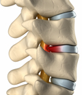
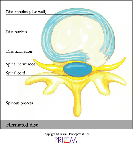
The spine is composed of many vertebrae stacked on top of each other. Between these bones are discs, which act as shock absorbers. The shock-absorbing discs resemble jelly donuts, each having a jelly-like center. As we age, the discs naturally become less flexible and more brittle. Normal disc degeneration, which naturally occurs with old age, can also cause pain. Discs can herniate in any direction--forward, centrally or, most commonly, backward and sideways in the direction of the spinal nerves.
Herniated discs account for a small percentage of back pain.
While herniated discs are often referred to as “slipped discs,” this really isn’t accurate because discs don’t ever slip out of position. They are actually attached by connective tissue to vertebrae above and below.
Click on image to enlarge
A disc herniation can be “contained” or “uncontained.” With a bulge, for example, the jelly center remains within the disc wall. "Uncontained" means the jelly center has broken through the annulus wall but stays connected to the nucleus pulposus. Or the herniation can be “sequestered,” when it breaks free from the nucleus and travels away from the disc.
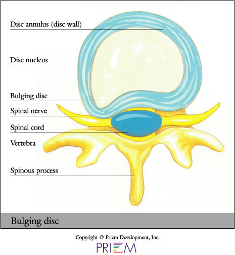
A bulging disc forms when the wall of
the disc is deformed but not necessarily herniated. The nucleus is
still contained in the wall. You NEVER need surgery to treat a bulging
disc.
[top]
Click on image to enlarge
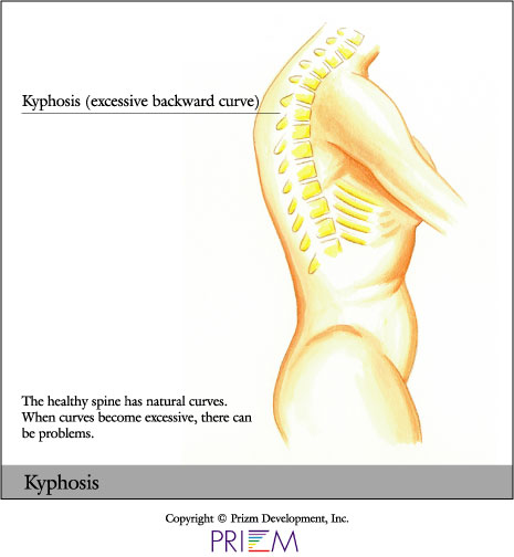
Kyphosis and lordosis are types of spinal deformities. While slight
curvature of the spine is normal and healthy, there are some cases
where it is over-pronounced and can cause both cosmetic deformity and
health risks. When the spine curves inward too much in the low back,
it is called lordosis. When the spine in the shoulder blade or mid-spine
area has too much forward curve, or too much of a hump, it is called
kyphosis. Kyphosis most often occurs in the thoracic area of the spine.
[top]
Spinal cord tumors are abnormal growths of tissue found inside the
bony spinal column, which is one of the primary components of the
central nervous system (CNS). Benign tumors are noncancerous, and
malignant tumors are cancerous. The CNS is housed within rigid, bony
quarters (i.e., the skull and spinal column), so any abnormal growth,
whether benign or malignant, can place pressure on sensitive tissues
and impair function. Tumors that originate in the brain or spinal
cord are called primary tumors.
[top]
Click on image to enlarge
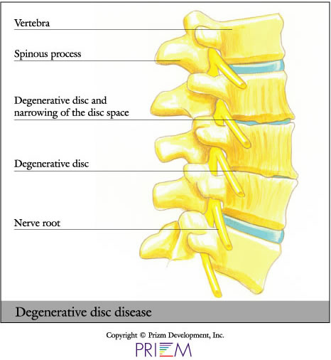
Degenerative disc disease commonly occurs with age as discs become more
brittle, less resilient and more prone to herniation. Degenerative disc
disease is the single most common diagnosis related to serious back and
neck pain. When a disc herniates in the spine, the surgeon can sometimes
simply remove a portion of the disc. In other cases, where the disc is
more damaged and must be removed, something must be placed into the disc
space. Otherwise, the two vertebrae will collapse on top of one another,
placing pressure on the nerve roots that branch off from the spinal cord.
[top]
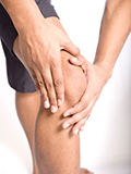
The back muscles provide support for the spine, allowing us to comfortably carry out our everyday activities. Back muscles can be grouped into three main categories. First, the extensor muscles allow us to stand up straight. Secondly, the flexor muscles allow us to bend forward. Finally, the oblique muscles enable us to rotate from side to side and keep everything stable and aligned.
If you think of the spine as a tall radio tower that
must withstand the force of crosswinds, the muscles and ligaments of
the back are the guy wires that provide support to the tower. The extensor
muscles enable us to arch our back and are located in the back. Flexor
muscles are also known as abdominal (stomach) muscles and are located
in front of the spine. The oblique muscles are located on our sides,
around the waist area, and they help stabilize our torsos and control
the pelvis.
[top]
Facet joints are the main "hinges" in our backs, allowing the muscles and vertebrae to move properly. Joints can lose their lubrication, swell and become painful, but if "well-oiled" with exercise and gentle stretching, joints will remain healthy.
[top]
We have made it easier to schedule appointments, click the button to fill out an online appointment request form or call our new central scheduling line 401-457-1500 to schedule an appointment.
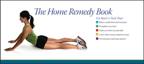
Have back or neck pain? Learn what causes symptoms and the home remedies that relieve pain. University Orthopedics mails out Home Remedy Books to residents throughout the New England area.
University Orthopedics in Providence, Rhode Island, is a regional referral center for patients with back and neck pain, joint pain, sports medicine problems, shoulder pain, hand problems, hip and knee pain, and foot and ankle injury. University Orthopedics utilizes fellowship-trained, sub-specialized physicians to treat a variety of orthopedic problems. Learn more.