- Locations
- Find a Physician
- By Physician
- By Department
- The Center for Spine Health
- Hand & Wrist Center
- Shoulder & Elbow Center
- Foot & Ankle Center
- Joint Replacement Center
- The Sports Medicine Center
- Pediatric Orthopedic Center
- Trauma & Fracture Center
- Osteoporosis and Bone Health
- Oncology Center
- Cartilage Repair Center
- Concussion Rehab Center
- OrthoDirect
- Careers
- Patient Portal
- Intranet
EOSedge™, a Next-Gen Imaging System Ideal for Pediatric and Adult Orthopedic Patients
The state-of-the-art imaging system with Flex Dose™ technology provides high-resolution images while minimizing radiation exposure
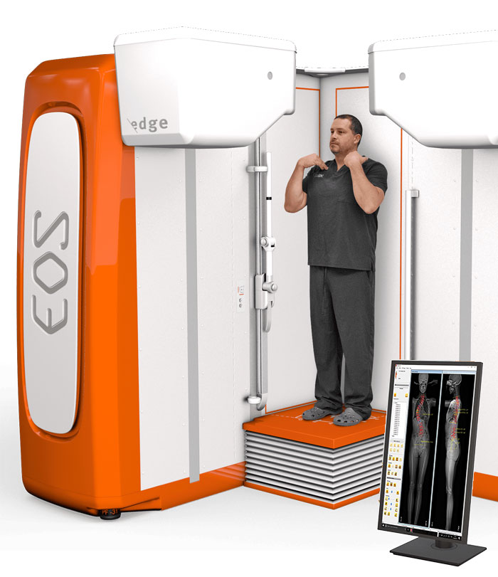
In an effort to continue our commitment to providing the highest quality care, University Orthopedics now offers EOSedge™, a Next-Gen Imaging System Ideal for Pediatric and Adult Orthopedic Patients.
What is EOS ®?
The EOS® system provides low dose, full body, images of patients in a weight-bearing, functional position. It is a bi-planar device that is based on two perpendicular X-ray beams that travel vertically while scanning the patient from head to toe. In a few seconds, the EOS exam produces two simultaneous frontal and lateral, low-dose images of the patient’s entire body without magnification or stitching.
Using the two full-body images, 3D models of your skeleton are able to be created. Your doctor will use the accurate measurements and data from the 3D model to make your diagnosis and prepare a personalized treatment for you as part of your shared decision making process.
EOSedge by EOS imaging
EOSedge is the latest technology by EOS imaging - which is already globally recognized for its low-dose, full-body biplanar imaging platform and its cloud-based digital spine ecosystem. This next-generation solution captures fully functional 2D biplanar images in seconds, using a high-resolution photon-counting detector for outstanding image quality for a broad range of patients. With the use of Flex Dose™ technology, EOSedge ups the ante in patient safety by substantially reducing radiation dose and minimizing the long-term impact to the patient.

Moreover, University Orthopedics will have access to a new web-based platform built around EOSedge images that uses artificial intelligence to evaluate a patient’s spine alignment, create surgical strategies based on patient-specific measurements, and reconcile surgical plans intraoperatively and postoperatively. This end-to-end platform will allow surgeons to visualize patient data, track trends, and create more predictable outcomes.
“The EOSedge system is a remarkable advancement in musculoskeletal imaging and it is our privilege to have this technology available to our surgeons and patients,” said University Orthopedics President Edward Akelman, MD. “By obtaining highly accurate patient data at a lower radiation dose, this new system enables our providers to make better informed clinical decisions while also prioritizing patient comfort and safety. We believe this signifies best-in-class care and a tremendous honor for us to be the first to offer EOSedge in New England.”
By modulating the radiation dose along the patient’s body - emitting less radiation along thinner parts of the patient and increasing it along thicker parts - Flex Dose delivers up to an 80% overall radiation reduction. Additionally, a Micro Dose function for follow-up exams can further reduce radiation exposure, delivering the equivalent amount of radiation received from living on Earth for one week. In addition, EOSedge’s open cabin allows medical teams to capture full-body images safely, quickly, and comfortably. Its accessible design enables easy entry and positioning for children, older patients, and patients with varying physical conditions.
Images from EOSedge seamlessly integrate with the EOS Insight digital platform, which has been available at University Orthopedics for several years.
Benefits for patients
Reduced radiation dose
The EOS system delivers 50%1 to 85%2 less radiation than traditional X-ray systems and 95%3 less dose than computed tomography (CT) scans. Reducing radiation dose is particularly beneficial for children requiring frequent imaging, such as children with spinal deformities like scoliosis. The Micro Dose feature further reduces radiation exposure, offering frontal and lateral pediatric full spine images at a dose that’s equivalent to only a week’s worth of natural radiation4.
High image quality in a weight-bearing position
Most imaging modalities capture images while you are lying down. With EOS, you will be standing or sitting during your exam. By capturing exams in an upright, weight-bearing position; physicians are able to better evaluate your global posture; understand the relationship between your spine, pelvis and lower limbs; as well as the compensatory mechanisms of the lower limbs.
This additional data is critical for physicians to improve diagnosis and treatment decisions and plan more precise surgical interventions, which can help to improve patient outcomes.
1. Diagnostic imaging of spinal deformities: reducing patient’s radiation dose with a new slot-scanning x-ray imager. Deschenes S et al. Spine. 2010
2. Comparison of radiation dose, patient comfort and financial break-even of standard digital radiography and a novel biplanar low-dose x-ray system for upright full-length lower limb and whole spine radiography. Dietrich TJ et al. Skeletal Radiol. 2013
3. Ionizing radiation doses during lower limb torsion and anteversion measurements by EOS stereoradiography and computed tomography. Delin C, et al. Eur J Radiol. 2013
4. EOS Micro Dose protocol for the radiological follow-up of adolescent idiopathic scoliosis. Ilharreborde B. et al. Eur Spine J. 2015
News
University Orthopedics First in New England to Offer EOSedge™, a Next-Gen Imaging System Ideal for Pediatric and Adult Orthopedic Patients

EAST PROVIDENCE, R.I. — Continuing its commitment to providing patients with cutting-edge care, University Orthopedics recently became the first practice in Rhode Island to add EOSedge to its suite of imaging tools. The use of this leading technology in radiographic imaging will help improve both patient safety and treatment outcomes.
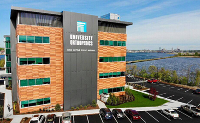
To celebrate the new technology, East Providence Mayor Robert DaSilva joined members of the University Orthopedics team and EOS representatives for a ribbon cutting this week at the practice’s Kettle Point location.
“Residents tell me all the time how happy they are that they don't have to travel far to get the care they need. They don't have to go over the bridge. They don't have to go to Providence or Boston. They come right here and get first-rate service,” Mayor DaSilva said. “And now our residents will have access to this cutting-edge technology that’s not available anywhere else in New England. That’s something to be proud of and it’s a testament to University Orthopedics and what they think about their patients.”
Click here to learn more.
Dr. Alan Daniels and Dr. Craig Eberson discuss the benefits of EOS imaging.
Do you Need an Appointment?
We have made it easier to schedule appointments, click the button to fill out an online appointment request form or call our new central scheduling line 401-457-1500 to schedule an appointment.
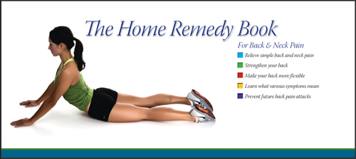
Home Remedy Book
Have back or neck pain? Learn what causes symptoms and the home remedies that relieve pain. University Orthopedics mails out Home Remedy Books to residents throughout the New England area.