- Locations
- Find a Physician
- By Physician
- By Department
- The Center for Spine Health
- Hand & Wrist Center
- Shoulder & Elbow Center
- Foot & Ankle Center
- Joint Replacement Center
- The Sports Medicine Center
- Pediatric Orthopedic Center
- Trauma & Fracture Center
- Osteoporosis and Bone Health
- Oncology Center
- Cartilage Repair Center
- Concussion Rehab Center
- OrthoDirect
- Careers
- Patient Portal
- Intranet
Spine Navigation and 3D Imaging
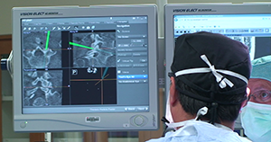 Most people who have watched a 3D movie on a big screen, will agree that it is a pretty cool technology. When it comes to 3D imaging for spine navigation to assist with complex spine surgeries, physicians and patients would both agree that this technology is a welcome addition to the operating room as well.
Spine navigation intra-operative guidance up close. Click image to watch video on computer assisted spine surgery technology. Video and Image used courtesy Stryker® Corporation. All rights reserved.
Computer assisted technology such as the Stryker® Spine Navigation System at University Orthopedics has been aiding orthopedic surgeons and neurosurgeons in many types of surgeries, such as knee or hip replacement and brain surgery, for years. The Spine Navigation System is an improvement on the fluoroscope, which provides a two-dimensional view during a surgical procedure. The Stryker Spine Navigation System offers doctors a comprehensive, fluid 3-D picture of the exact surgical site in the patient — an exact roadmap of the patient’s spine.
Dr. Craig Eberson and Dr. Phillip Lucas at the University Orthopedics Spine Center in Rhode Island now apply the power of navigation technology to spine surgery. The Spine Navigation System places the spine surgeons at University Orthopedics at the forefront of computer assisted surgery for spine in the nation.
The University Orthopedics Spine Center already receives referrals from across Southern New England. These patients with complex spinal problems travel great distances to University Orthopedics in search of pain relief and for help in getting their lives back. This 3D surgery technology allows spine surgeons like Dr. Eberson and Dr. Lucas to perform complex spine surgeries with improved safety, speed and precision.
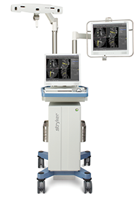
Spine Navigation System. Image
courtesy of Stryker Corporation.
All rights reserved.
How does the technology work?
Before surgery, the patient will have a CT scan. The images are then downloaded to the navigation computer. Software is used to develop a virtual, 3D model of the spine. The spine surgeon inserts a special Stryker probe into the patient through a small incision. The probe transmits a signal back to the navigation system. The software uses the patients CT Scan with information from the Stryker probe to transmit a real-time view of where the instruments are throughout the procedure.
Benefits of Spine navigation and 3D imaging:
- Helps reduce the radiation exposure of both the patient and medical staff as it eliminates the need for multiple X-ray images.
- Offers the surgeon a complete picture of patient anatomy prior to surgery. This allows the surgeon to better prepare for surgery and can result in shorter surgery times.
- Surgeons have real-time knowledge of instruments and implant position during surgery.
- Reduces potential for incorrect implant placement during surgery.
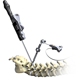
Spine Model and Smart Instrument. Image
courtesy of Stryker Corporation.
All rights reserved.
The navigation technology does not replace the surgeons skills. “Using the Spine Navigation System during spine surgery allows the surgeon to be more precise. As an example, before this technology, surgeons were dependant upon 2D images, their grasp of anatomy and their existing knowledge of the patient to determine where to insert pedicle screws to fuse vertebrae in the lumbar spine,” explains Dr. Daniels. “With the Stryker Spine Navigation System and 3D images during surgery, spine surgeons are even more exact.”
The use of image-guidance technology in all types of spinal surgery is rapidly growing. Spine fusion surgeries that help relieve pain resulting from injury, degenerative disc disease, spinal curvatures or arthritis are the most common navigated surgeries. After spine fusion surgeries, many patients can have significant reduction in pain and a successful return to an active lifestyle.
The University Orthopedics Spine Center is pleased to incorporate the Spine Navigation System to aid in complex spine surgeries to treat disc herniations, spinal stenosis, spinal tumors, trauma and scoliosis.
About The Center for Spine Health at University Orthopedics
The Center for Spine Health at University Orthopedics includes a team of physicians, nurse practitioners and physical therapists working together to provide comprehensive treatment of spinal disorders in adults and children. Our clinical team has specialized expertise in the management of spinal pain, disc herniations, stenosis, tumors, trauma and scoliosis (abnormal curvature of the spine).
To make an appointment with the University Orthopedics Spine Center, please call (401) 414-BACK (2225).


